INTRODUCTION
The family Rhodomelaceae includes 1,080 species classified in 158 genera (Guiry and Guiry 2019), and exhibit an extraordinary morphological diversity (Womersley 2003, Díaz-Tapia et al. 2017). Usually, members of this family are easily recognized by their thalli consisting on an axial filament whose cells are surrounded by pericentral cells and by the sympodial branching pattern (Womersley 2003). Moreover, most species in the Rhodomelaceae have trichoblasts, apical monosiphonous branches that are usually unpigmented (Maggs and Hommersand 1993, Womersley 2003).
The Rhodomelaceae is the most diverse family of the red algae, and the number of recognized species is continuously growing. The discovery of new species is often related to the finding of cryptic diversity, both when comparing molecular data of specimens from distant locations or from the same region (e.g., Zuccarello et al. 2015, Muangmai et al. 2016, Savoie and Saunders 2016, Díaz-Tapia et al. 2018, Schneider et al. 2018). More surprisingly, some species with conspicuous morphological differences regarding other members of the family remained unnoticed. One of the causes is that they grow in poorly explored habitats, such as the deep subtidal (Bárbara et al. 2013, Kim and Kim 2014). Intertidal sand-covered rocks are other common, more accessible, but often poorly explored habitat. The presence of sediments in intertidal rocks negatively affects many benthic organisms, and sand-covered rocks host a particular algal assemblage (Airoldi 2003, Díaz-Tapia et al. 2013a) in which several new red algal species have been recently described (Díaz-Tapia and Bárbara 2013, Díaz-Tapia et al. 2013b, D’Archino et al. 2015). During our surveys of marine red algae in this habitat along the coasts of Espírito Santo and Rio de Janeiro (southeastern Brazil), we found abundant populations of a rhodomelacean species consisting of large plants (up to 25 cm in length) with seven pericentral cells and a dense cortication from close to the apices. The morphology of this conspicuous species does not match with the species previously recorded in the region. Even its generic assignment was uncertain, as these characters resemble to some genera of the tribes Chondrieae, Alsidieae or Pterosiphonieae. The objective of this work is to clarify the taxonomic identity and phylogenetic relationships of the Brazilian species using molecular (rbcL and cox1 genes) and morphological data.
MATERIALS AND METHODS
Plants of the targeted species were collected between 1986 and 2017 in Espírito Santo (21°03′ S, 40°52′ W to 19°52′ S, 40°03′ W) and Rio de Janeiro (Praia Rasa; 22°44′ S, 41°57′ W) from the intertidal zone or in the drift (see material examined and Supplementary Table S1). Samples preserved in silica gel desiccant, collected in 2006 and 2014, were used for DNA extraction. Plants for morphological studies were fixed in 4% formalin in seawater. Sections for anatomical studies were made by hand using a razor blade and stained in 1% aqueous aniline blue acidified with 1 N HCl. Sections were photographed on Ilford 50 ASA film (Harman Technology Ltd., Cheshire, UK) with an Olympus BH-2 photomicroscope (Olympus Corporation, Tokyo, Japan). Representative material was deposited in the SP (Institute of Botany) herbarium, São Paulo, Brazil.
DNA was extracted from silica gel-dried material following Saunders and McDevit (2012). Polymerase chain reaction (PCR) amplification was carried out for rbcL using the primers F2/R1452, F7/RrbcsStart, F7/R893 or F57/rbcLrevNEW (Freshwater and Rueness 1994, Mamoozadeh and Freshwater 2011, Saunders and Moore 2013, Díaz-Tapia et al. 2018) and for cox1 using the primers GwsFn/Cox1R1 (Saunders 2008, Le Gall and Saunders 2010) or GazF1/GazR1 (Saunders 2005). Reactions were performed in a total volume of 25 μL, consisting of 5 μL 5× MyTaq reaction buffer, 0.7 μL 10 μM of forward and reverse primers, 0.125 μL 1 U/μL My Taq DNA Polymerase (Bioline, London, UK), 17.475 μL MilliQ water and 1 μL template DNA. The PCR profile consisted of initial denaturation (93°C for 3 min), 35 cycles of denaturation (94°C for 30 s), primer annealing (45°C for 30 s), and extension (74°C for 90 s) and final extension (74°C for 5 min). The PCR products were purified and sequenced commercially by Macrogen (Seoul, Korea).
Two sequences were generated in this study for the targeted species for each gene (rbcL and cox1), and blast search in GenBank for the rbcL sequences of the Brazilian unidentified species indicated Alsidium seaforthii (Turner) J. Agardh, in the tribe Alsidieae, was that the most similar species (similarity 97.23%). Therefore, we downloaded from GenBank all the available rbcL and cox1 sequences for species of the tribe Alsidieae. We also determined five and ten new rbcL and cox1 sequences for species of the genus Alsidium C. Agardh from Brazil, the Canary Islands and the Mediterranean Sea (Supplementary Table S1). We selected 17 sequences, one per haplotype, for the rbcL and cox1 phylogenetic analysis. When several sequences were available for a haplotype, we selected the largest. Sequences and their corresponding GenBank accession numbers are listed in Supplementary Table S1. Sequences were aligned using Muscle in Geneious 6.1.8 (Kearse et al. 2012). Phylogenetic trees for rbcL and cox1 were estimated with maximum likelihood (ML) using RAxML 8.1.X (Stamatakis 2014). GTR-Gamma was used as the nucleotide model; branch support was estimated with 100 bootstrap replicates. The genera Chondria C. Agardh and Acanthophora J. V. Lamouroux (tribe Chondrieae) were selected as outgroup for the rbcL and cox1 trees, respectively. Our outgroup selection was based on the phylogenomic analyses of the major lineages of the Rhodomelaceae that resolved a clade formed by the Chondrieae and Laurencieae as sister to the Alsidieae (Díaz-Tapia et al. 2017).
RESULTS
Phylogeny
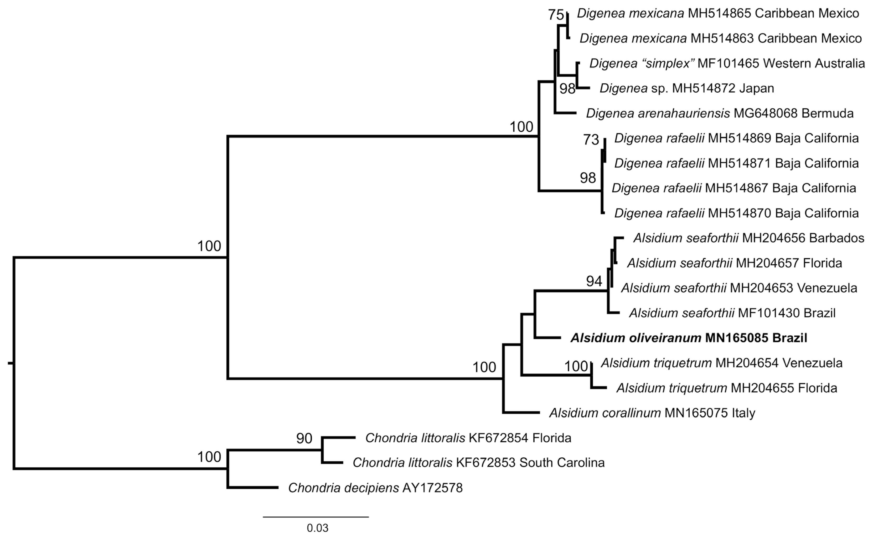
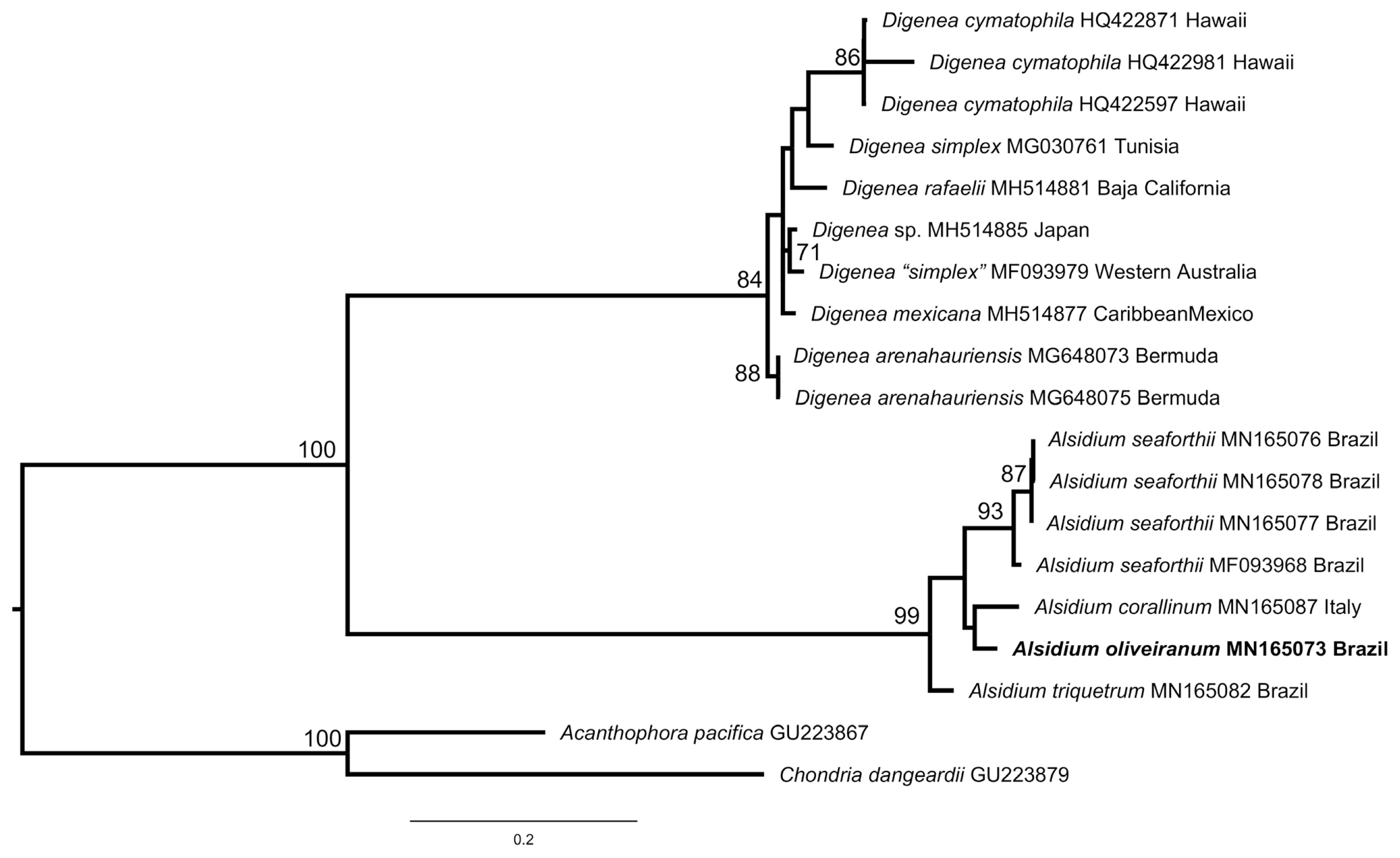
The RAxML phylogenetic analysis of rbcL sequences (Fig. 1) placed a unique Brazilian species of Alsidium in a fully supported clade that included other species of the genus. Within this clade the Brazilian species was placed as sister to A. seaforthii, but this relationship was unsupported. The Alsidium clade was sister to the fully supported Digenea C. Agardh clade. The cox1 tree (Fig. 2) had a similar topology than the rbcL tree. In the cox1 tree, the Brazilian species was placed as sister to the generitype Alsidium corallinum C. Agardh, but again, this relationship was unsupported.
Two rbcL and cox1 sequences for the new Brazilian Alsidium were identical. Seven rbcL sequences for A. corallinum from the Mediterranean Sea (three) and the Canary Islands (four) were identical. Likewise, four cox1 sequences of A. triquetrum (S. G. Gmelin) Trevisan, from Mexico and Brazil, were identical. A. seaforthii from Brazil was genetically more diverse in the cox1 gene, and four haplotypes that diverged by 0.1–2% (1–14 bp) were discovered for the species. Sequence divergence between the Brazilian species and other species of the genus Alsidium, was ≥2.5 and 4.2% in the rbcL and cox1 genes, respectively. These findings allow for the description of a new species of Alsidium from Brazil.
Morphological observations
Alsidium oliveiranum S. M. Guimarães & M. T. Fujii sp. nov. (Figs 3–5)
Diagnosis Thallus consisting of a basal crust, from which many cylindrical, erect branches are formed. Erect branches up to 1 mm in diameter, with seven pericentral cells and heavily corticated from close to the apices. Vegetative trichoblasts scarcely developed if present. Reproductive structures formed on clustered determinate lateral endogenous branches. Spermatangial branches formed on modified trichoblasts and replacing them, consisting of thin, flat discs, lacking marginal sterile cells. Cystocarps globose. Tetrasporangia spirally arranged on fertile branches, one per segment.
Holotype SP470454, Sep 10, 2014, M. T. Fujii & P. Díaz-Tapia, sand-covered rocks in the low intertidal.
Type locality Praia da Cruz; Marataízes, Espírito Santo, Brazil.
Etymology Named in honor of Dr. Eurico Cabral de Oliveira F°, from the University of São Paulo, for his contributions to our understanding of the Ceramiales from Espírito Santo, Brazil.
Molecular vouchers MN165085 rbcL, MN165073 cox1.
Other specimens examined Espírito Santo: Aracruz County, Praia dos Padres, Oct 22, 1996, S. M. P. B. Guimarães & M. T. Fujii, SP470448. Serra County, Praia da Baleia, Sep 17, 1986, S. M. P. B. Guimarães, SP470447. Anchieta County, Praia de Parati, Jul 20, 1997, S. M. P. B. Guimarães & M. T. Fujii, SP470449; Oct 4, 2006, M. T. Fujii, SP470451; Sep 8, 2014, M. T. Fujii, SP470452. Anchieta County, Ponta dos Castelhanos, Sep 9, 2014, M. T. Fujii, SP470453. Itapemirim County, Praia de Itaoca, Oct 5, 2017, L. P. Soares, SP470455. Marataízes County, Praia da Cruz, Sep 10, 2014, M. T. Fujii, SP470454; Praia de Marataízes, Sep 15, 2001, S. M. P. B. Guimarães & M. T. Fujii, SP470450. Rio de Janeiro: Armação dos Búzios Country, Praia Rasa, Oct 24, 2011, M. T. Fujii, SP470467.
Habitat and distribution Plants grow up in tufts forming dense intertidal populations in Espírito Santo and Rio de Janeiro, southeastern Brazil (ca. 20° S), growing on sandstone beach reefs or on rocky outcrops often buried by sand and subjected to moderate water movement. Plants were also collected in the drift.
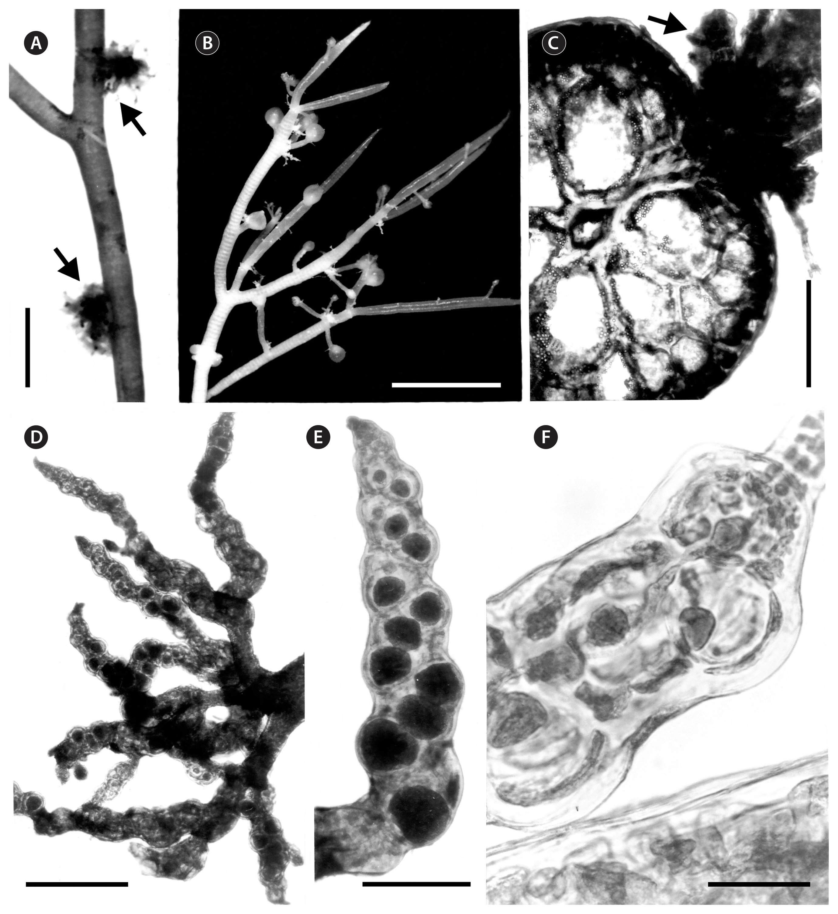
Vegetative morphology Thalli consisted of a basal crust 2–3 cm in diameter bearing tufted erect axes up to 25 cm high and 1 mm in diameter (Fig. 3A & B). Erect axes terete, scarcely branched alternately to irregular, and bearing clusters of short determinate endogenous lateral branches. Thallus not adhering to herbarium paper when dried; rigid and red to reddish-brown or wine-red in color. Erect axes with polysiphonous structures and heavily corticated. Apical cell of erect axes dome-shaped, 17–25 μm in diameter, dividing transversely forming axial cells which divided longitudinally producing the pericentral cells (Fig. 3C). Branches of vegetative axes formed endogenously close to the apices (Fig. 3G). Cortication developed from close to the apices, initially formed by divisions at the margins of the pericentral cells. Later, the cortical cells formed a continuous layer, so that the pericentral cells were only visible in surface view at the apices. In surface view, cortical cells in middle portions of thalli were rounded to polygonal, 15–55 × 17–98 μm (Fig. 3D). In cross-section (Fig. 3E), thallus consisted of an axial cell, seven rounded to polygonal pericentral cells, 123–250 μm in diameter, a layer of medullary unpigmented rounded to polygonal cells, 115–150 μm in diameter, and an outer layer of pigmented and rounded to rectangular cortical cells, 27–100 μm in diameter. Secondary pit connections formed between adjacent outer cortical cells. In longitudinal section, pericentral cells were rounded to polygonal, with the same length as axial cells (Fig. 3F). Lateral endogenous determinate branches scarce, forming clusters. Trichoblasts rare, formed at the apices of young vegetative branches when present; short, to 140 μm long, and dichotomously branched 1–2 times (Fig. 3G–I).
Reproductive morphology Reproductive structures produced on lateral endogenous branches (Fig. 4A–D). Spermatangial branches consisted of thin, flat discs, lacking sterile marginal cells and replacing trichoblasts. In fertile female plants, procarps formed on modified trichoblasts on the adaxial side of short determinate laterals in a spiral pattern (Fig. 5A & B). Few short vegetative and reproductive trichoblasts present on young female branchlets (Fig. 5D & E). The entire pericarp seems to develop from the third and fourth pericentral cells of the fertile segment (Fig. 5C). Mature cystocarps globose, c. 1 mm in diameter, corticated, slightly flat at the top and with an apical ostiole (Figs 4B, 5F & G). Owing to its growth, mature cystocarps become distal on a short lateral branchlets. Apical portions of branchlets can be observed as ligulate appendices (Fig. 5F). Tetrasporangial branches originated endogenously from axial cells and densely clustered (Fig. 4A, C & D), corticated, cylindrical with tapered tips, 1.0–2.0 mm in length and 100–200 μm in diameter (Fig. 4E), distributed sparsely along the thallus. Tetrasporangia 90–100 μm in diameter formed adaxially from pericentral cells. One tetrasporangium occurred in each segment in a spiral sequence, causing swelling on the side where the spores are borne (Fig. 4E & F). Mature tetrasporangia covered by two presporangial cover cells and elongated cortical cells.
DISCUSSION
The Brazilian species in our phylogeny nested in a clade with species of the genus Alsidium. The most distinctive morphological characters at the generic level observed in this species were as follows: (1) the thallus consists of a basal crust that produces erect axes; (2) the erect axes are cartilaginous with seven pericentral cells and are corticated beginning close to the apices; (3) spermatangial branches replace the trichoblasts and are plate-like; (4) one tetrasporangia per segment is formed on endogenous determinate branches. All of these characters are in agreement with the current delineation of the genus Alsidium (García-Soto and Lopez-Bautista 2018). Moreover, the new species is characterized by a development of the cortex that differs from Digenea, the other genus in the tribe Alsidieae. In A. oliveiranum, the formation of the cortex commences close to the apices and the first cortical cells are divided from the margins of the pericentral cells. Later, the cortex continues its development forming a continuous layer that covers the pericentral cells. Cortical development in A. oliveiranum is similar to that observed in other corticated members of the genus (A. seaforthii and A. corallinum) (PD personal observation). The development of the cortex in Digenea, by contrast, is unusual compared with Alsidium and other Rhodomelaceae. The pericentral cells initially divide into discrete packets of cortical cells that cover each of the pericentral cells (Falkenberg 1901, Norris 1994, Boo et al. 2018, Schneider et al. 2018). Such pattern is conspicuous in the apical parts of Digenea cymatophila (R. E. Norris) Díaz-Tapia & Maggs as well as in the determinate branches of other species of Digenea (Falkenberg 1901, Norris 1994, Boo et al. 2018, Schneider et al. 2018). Later, the cortex of Digenea further develops obscuring this pattern in older parts of the thallus. Therefore, the cortical development is a useful character for delineating Alsidium from Digenea. Most species in Digenea share a common and distinctive habit, as the main axes are densely clothed with short determinate branches. However, the habit of D. cymatophila, with scarce determinate branches, differs from other congeners and resembles Alsidium. In fact, D. cymatophila was originally assigned to Alsidium (Norris 1994) and later transferred to Digenea based on its placement in phylogenetic analyses (Díaz-Tapia et al. 2017, Fig. 2 in this work). A. oliveiranum is particularly similar in outline morphology to D. cymatophila, as both species consist of a basal crust from which scarcely branched erect axes arose, and determinate branches are clustered and mainly formed in relation to reproductive structures. The cortex development is particularly useful for the generic assignment of this pair of species.
Alsidium oliveiranum clearly differs from the other three species of this genus that have been molecularly characterized (A. triquetrum, A. seaforthii, and A. corallinum) by sequence divergence ≥2.5 and 4.2% in the rbcL and cox1 genes, respectively. Moreover, they can be distinguished by their morphological characters (Table 1). A. triquetrum and A. seaforthii, formerly included in the genus Bryothamnion Kützing, have compressed or triangular thallus differing from the terete axes in A. oliveiranum (Littler and Littler 2000, García-Soto and Lopez-Bautista 2018). Likewise, A. corallinum and A. oliveiranum differ morphologically in several characters: (1) axes have 5–8 vs. 7 pericentral cells; (2) the thallus being clothed with abundant determinate branches vs. determinate branches being scarce and mainly produced in relation to reproductive structures; and (3) tetrasporangia forming in non-clustered vs. clustered lateral determinate branches (Kützing 1865, Rodríguez-Prieto et al. 2013).
Other four species are currently recognized in the genus Alsidium (Guiry and Guiry 2019), but molecular data are unavailable at present. Alsidium helminthochorton (Schwendimann) Kützing was originally described and is endemic to the Mediterranean Sea (Kützing 1865, Rodríguez-Prieto et al. 2013). Relevant differences between A. helminthochorton and A. oliveiranum include: (1) axes with 7–9 vs. 7 pericentral cells; and (2) the attachment by prostrate axes vs. a basal crust (Kützing 1865, Rodríguez-Prieto et al. 2013). Two very small species that differ in the number of pericentral cells, Alsidium pacificum E. Y. Dawson and Alsidium pusillum E. Y. Dawson, were described in the Palmyra Atoll and the Galapagos Islands, respectively (Dawson 1959, 1963), and were considered conspecific by Norris (1994). Their short erect thalli (up to 15 mm in length) contrast with the long thalli in A. oliveiranum (up to 25 cm). Moreover, they have a system of prostrate axes that differs from the basal crust in A. oliveiranum (Dawson 1959, 1963). Finally, Alsidium vagum (Zanardini) Zanardini was described from the Mediterranean Sea and was not recorded after its original description (Guiry and Guiry 2019). Such description did not provide morphological details enough to ascertain its identity and further studies are required to clarify its taxonomy (Zanardini 1851, Bompard 1867). Therefore, our molecular and morphological studies clearly distinguish the Brazilian species from previously described species in the genus Alsidium and consequently we erected the new species A. oliveiranum here.
In Brazil, two species of Alsidium were traditionally reported based on morphological studies: A. seaforthii and A. triquetrum. Both are distributed from the Atlantic North America to Brazil (Guiry and Guiry 2019). A. oliveiranum has a much narrower distribution and is endemic to the states of Espírito Santo and Rio de Janeiro; despite our sampling efforts, it was not found elsewhere. In Brazil, A. seaforthii has the widest range, extending southward to the coast of São Paulo, while A. triquetrum is restricted to the northeastern coast (up to Bahia). A. oliveiranum and A. seaforthii occur sympatrically on the Espírito Santo and Rio de Janeiro (Guimarães 2006). This result is unsurprising, as this region hosts a particularly high algal species diversity, including rare and endemic species (De Oliveira Filho 1969, Guimarães and Fujii 1998, Chen et al. 2019). Moreover, its seaweed diversity is continuously growing as the use of molecular assisted identification is contributing to the discovery of new species (Nauer et al. 2015, Iha et al. 2016, Ximenes et al. 2017, Brunelli et al. 2019a, 2019b). The high diversity in this peculiar coast has been attributed to the substrate heterogeneity and availability, as well as the particular seawater temperature conditions because this region is a transition zone between tropical and subtropical regions and it is affected by upwelling events (Guimarães 2003).
Our finding contributes to better understand the diversity of the genus Alsidium and the tribe Alsidieae. This tribe, with 15 recognized species, has a reduced species diversity compared with other tribes in the Rhodomelaceae and most species have restricted distributions in tropical and subtropical coasts: the Mediterranean and Macaronesian Islands (A. corallinum and A. helminthochorton), Pacific Islands (A. pusillum, A. pacificum, and Digenea cymathophyla), the Pacific and Atlantic coasts of the Americas (A. seaforthii, A. triquetrum, “Bryothamnion” pacificum W. R. Taylor) and South Africa (D. subarticulata Simons) (Guiry and Guiry 2019). Digenea simplex (Wulfen) C. Agardh is the only species of the tribe that has been widely reported in tropical and subtropical coasts worldwide (Guiry and Guiry 2019). However, the sequencing of material from the type locality, the Mediterranean Sea, led to the discovery of cryptic diversity within what had been referred to as D. simplex in the Americas where three new species have been segregated recently from D. simplex (Boo et al. 2018, Schneider et al. 2018). Interestingly, the Atlantic and Pacific coasts of America, with six currently recognized species, host the largest diversity of species of the tribe Alsidieae.
In conclusion, in this paper we described a new species of Alsidium endemic to Brazil, a species that was previously overlooked because of the lack of study of its intertidal habitat on sand-covered rocks. Interestingly, this is also the typical habitat for other species of the tribe Alsidieae (e.g., Boo et al. 2018, Schneider et al. 2018). Our work contributes to better understand the species and morphological diversity of the tribe Alsidieae.